Applied methods
- In vivo methods:
- synchronization of ovulation in cows and pigs, superovulation and insemination,
- ultrasound diagnostic (USG) of reproductive organs in cows, pigs and mares,
- laparotomy of cows and pigs (blood vessels cannulation in reproduction system, corpus luteum microdialysis and other structure),
- intravital acquisition ovaries and corpus luteum (laparotomy and calpotomy),
- uterine mucous membrane biopsies in cows;
- In vitro methods:
Enzymatic isolation and cells culture:
- epithelium and stromal cells from the endometrium of cows, pigs, mares and cats,
- steroidogenic cells from the corpus luteum of cows, mares and cats,
- granulosa cells from the ovarian follicle of cows,
- endothelium cells of corpus luteum and mucous membrane blood vessels in cows, pigs, mares and cats,
- epithelial cells of bovine mammary gland cisternae,
- culturing adherent and suspension cell lines,
- transfection of cells;
Tissues incubation:
- cows, pigs, mares and cats endometrium explants,
- cows, pigs and mares corpus luteum explants,
- bovine mammary gland cisternae explants;
Virology methods:
- isolation, concentration and preparation of viral stocks,
- infectivity assay (determining MOI).
- Analytic methods
3.1. Molecular biology methods:
- pro- and anti apoptotic factor (TNFα, IFNγ, IL-1α, Il-6, FasL, BCL2, BAX, Caspase3, Caspase8) expression and their receptors,
- enzymes steroidogenic pathway (3β-HSD, StAR, CYTp450)expression,
- enzymes of arachidonic acid metabolism pathway expression:
- leukotrienes pathway (5-LOX) and their receptors,
- prostaglandins pathway (PTGS-2, PGES, PGFS, 9KPGR, AKR1B5),
- oksitocin and their receptors expression,
- nerves growth factor expression and its receptors (TrkA, p75) with used Real Time PCR and semiquantitative RT PCR for analysis of mRNA levels and Western Blot method for analysis of protein level,
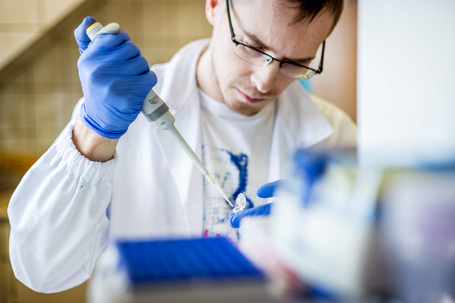
- cloning/ PCR (eg. viral genes),
- Western blot analysis (eg. expression level of viral latent and lytic proteins),
- Southern blot,
- Electromobility Shift Assay (EMSA) eg. binding of viral protein to sequences in viral genome;
3.2.Radioimmunoassay (RIA) method of hormones:
- protein hormones (LH, PRL) and their receptors (LH/hCG) with iodine marked specimen used (J-125),
- steroidogenic hormones (P4, A4, T, E1, E2, cortisol), prostaglandins and methabolit of PGF2α – PGFM, with tritium marked hormones (H3) and protein hormones iodination (LH, PRL) and cytokine (IL-1β, IL-6, TNF-α);
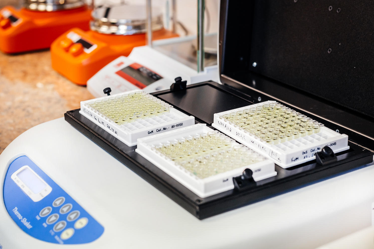
3.3. Immunoenzymatic (ELISA) method:
- protein hormones (LH),
- steroidogenic hormones (P4, T4, E2, cortisol),
- prostaglandins (PGE2, PGF2α),
- oxytocin and endothelin with hormones marked horseradish peroxidase (HRP) or biotin used;
3.4. Immunocytochemistry localization:
- arachidonic acid pathway enzymes in ovary, uterus and epithelial cells of mammary gland cisternae (PTGS-2, PGES, PGFS, AKR1B5, 5-LO),
- cytokine in ovary and uterus (TNF-α, IL-1α, IL-1β and others),
- enzymes of cholesterol pathway in ovary (P450scc, 3β-HSD, aromatase) and epithelial cells of mammary gland cisternae (11β-HSD, 3β-HSD),
- receptors (TNFR I, TNFR II, IL-1αR, IL-1 βR, LTRI, LTRII, GC-R, LPAR-1, LPAR-2, LPAR-3, LPAR-4, LPAR4, OTR, TrkA, p75),
- neurotransmitters and their markers (NPY, VIP, SOM, SP, GAL, CGRP, PACAP, DβH, VACHT, nNOS),
- fluorescence in situ hybridization (FISH) – detection of viral genome in the cell,
- immunofluorescence staining (IFA) of viral proteins in infected cells also combined with chromosome spreads,
- fluorescence activated cell sorting (FACS) analysis – analysing cell cycle;
3.5. Critical parameters and blood gasometry analysis (RAPIDpoint 405 SIEMENS);
3.6. Morphology blood analysis (ADVIA 2120i SIEMENS);
3.7. Chemiluminescent immunochemical blood analysis (IMMULITE®1000 SIEMENS),
Equipment
- Two workplaces for isolation and cells cultured:
- Laminair chamber Thermo Scientific MSC-ADVANTAGE (180)
- Laminair chamber Polon’120
- Incubator CO2 ThermoForma
- Laminair chamber Esico Class II BSC
- Incubator CO2 Sanyo
- Incubator Memmert IMCO2-153
- Centrifuge MPW 350R – 2
- Centrifuge MPW 223E
- Centrifuge Eppendorf 5804R
- Peristaltic pump VederFlex Scientific
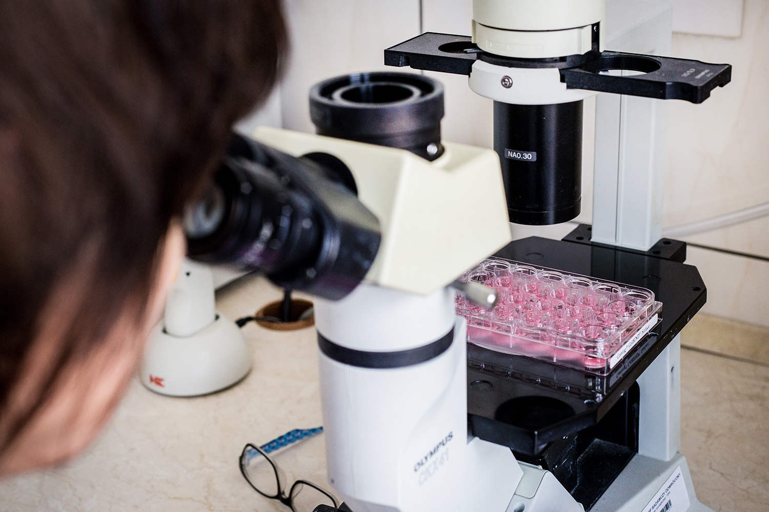
- Inverted microscope (standard) Olympus CKX41 – 2
- Microscop Olympus CX31RBSF-2
- Incubator with shaker EB TH15(Edmund Bühler) – 2
- Incubator with shaker Major Science SWB-10L-1
- Water bath with shaker Julabo SW23
- Ultrasonic pipette-washer Sonore
- Autoclave Sanyo
- Incubator Sanyo MOV-112F
- Dryer Binder E28
- Dryer Terma
- Dishwasher Smeg GW1060C
- Workplace of nucleic acids isolation:
- Laminair chamber Holten 90
- Centrifuge Hettich Universal 32R
- Centrifuge Eppendorf miniSpin
- Gel electrophoresis device BIO-RAD PowerPac
- UV Ttransilluminator WEALTEC
- Thermocycler BIO-RAD MJ Mini
- Whipper TissueRuptor Qiagen
- Centrifuge lab Waeltec E-Centrifuge
- Water bath with shaker Major Science MW-WB
- Vortex BioRad Sproud
- Vortex Mixer LabNet
- Western blot workplace:
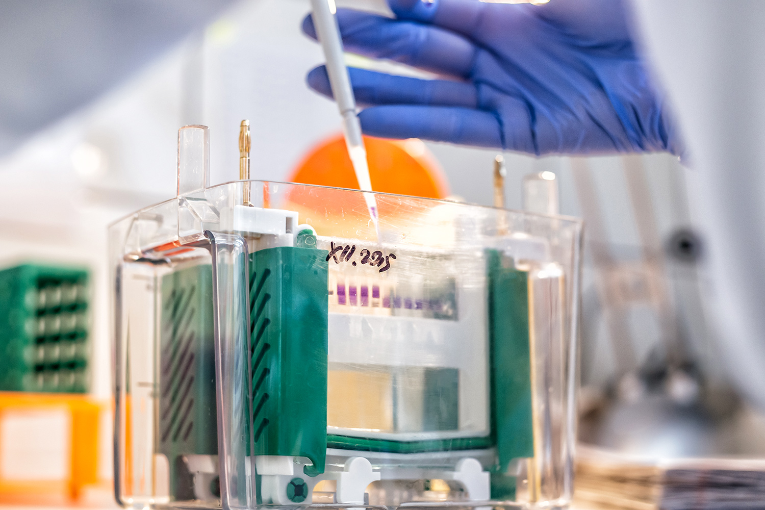
- Polyacrylamide gel electrophoresis device BIO-RAD PowerPac,
- Semi-dry transfer system TE70PWR Amersham Biosciences,
- Wet transfer system BIO-RAD PowerPac,
- RollMixer Chemland,
- Protein detection system SNAP i.d. Millipore;
- Integrated Immunodiagnostic Laboratory
- Laboratory of Molecular Microbiology and Virology
5.1 Cell culture and GMM class II workstation
5.2 Workstation for isolation of fibroblasts from human tissue
5.3 Workstation for protein expression analysis (SDS PAGE + Western blot)
5.4 Workstation for solid and liquid bacterial culture and for co-culture of bacteria with tissues.
